Cystography
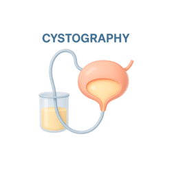
A Cystography (or cystogram) is an X-ray study of the bladder and urethra. A contrast dye is gently placed into the bladder so the urinary system shows up clearly on the images.
This page walks you through the Cystography and VCUG procedures in simple language—what they are, how to prepare, what happens during the test, and how doctors interpret VUR grades.
Two related imaging tests that help your care team see how your bladder and urethra are working and whether urine is flowing in the right direction.
A Cystography (or cystogram) is an X-ray study of the bladder and urethra. A contrast dye is gently placed into the bladder so the urinary system shows up clearly on the images.
Voiding Cystourethrography (VCUG) is a specialized cystography performed while you empty your bladder. It shows how urine moves and whether any flows backward toward the kidneys.
VCUG is the gold-standard test for Vesicoureteral Reflux (VUR), a condition where urine travels backward from the bladder up toward the kidneys instead of out of the body.
To evaluate the bladder and urethra, doctors use an imaging test called cystography. A small catheter is placed into the bladder, contrast dye is instilled, and a series of X-ray images are taken. This lets the radiologist see the shape of the bladder, the urethra, and how well everything is working.
A Voiding Cystourethrography (VCUG) is the most common form of cystography. After the bladder is filled with contrast, images are captured while you urinate. This helps your care team look for vesicoureteral reflux (VUR)—urine flowing backward toward the kidneys instead of in the normal direction. Understanding what will happen and why the test is ordered can make the experience less stressful for you or your child.
Good preparation keeps the visit smoother and helps your team capture clear, useful images.
Most people do not need special fasting before a cystography or VCUG, but a few simple steps make a big difference. Use this checklist to get ready and follow any extra instructions from your imaging center.
For age-specific tips, see the dedicated pediatric guide: VCUG preparation for children.
A two-part process: filling the bladder with contrast and then capturing images as you empty.
The procedure begins with careful placement of a small urinary catheter through the urethra into the bladder. This step can feel uncomfortable or briefly stinging, but staff work slowly and explain each part.
Once the catheter is in place, contrast dye flows into the bladder. As the bladder fills, the technologist takes X-ray images to study the bladder’s shape and check for structural concerns such as diverticula, fistulas, or abnormal outlines.
When your bladder is full, you will be asked to empty it while standing, sitting, or lying—depending on the setup and age of the patient. During this time, the radiologist captures rapid images of the contrast as it leaves the bladder.
This is the key part of Voiding Cystourethrography (VCUG). The images reveal whether contrast flows in the right direction or backward toward the kidneys, which would suggest Vesicoureteral Reflux (VUR).
Worried about radiation exposure? Learn more in: Radiation safety in diagnostic imaging.
What radiologists look for, how VUR is graded, and what those grades usually mean for treatment decisions.
After the images are taken, the radiologist reviews how the bladder fills and empties, the outline of the bladder and urethra, and whether any contrast moves backward toward the kidneys. When VUR is present, it is usually graded on a five-point scale from I (mild) to V (severe).
Mild grades might only need monitoring and preventive antibiotics. Higher grades, especially in children with repeated infections or kidney changes, may call for closer follow-up or surgical consultation. Your doctor will explain which findings apply to you or your child and how they connect to overall kidney health.
For more technical details, health professionals often refer to: NIDDK information on VUR (external resource) .
VUR grades help your care team match treatment intensity to the severity of backward flow and kidney involvement.
When VCUG confirms VUR, the radiologist chooses a grade between I and V. Lower grades often improve over time, especially in children, while higher grades can require more active treatment. This table summarizes common descriptions and typical management approaches; always rely on your own clinician’s advice for personal decisions.
| Grade | What the images show | Typical treatment approach |
|---|---|---|
| Grade I | Reflux into the ureter only, without reaching the renal pelvis (the area that collects urine in the kidney). | Observation and sometimes low-dose antibiotics, especially in children with infections. |
| Grade II | Reflux up to the renal pelvis but no widening (dilation) of the ureter or collecting system. | Antibiotic prophylaxis and regular monitoring; many children outgrow lower grades over time. |
| Grade III | Mild to moderate dilation of the ureter and collecting system while the inner edges of the kidney cups remain relatively sharp. | Closer follow-up, medication, and periodic imaging; in some cases, surgical options are discussed. |
| Grade IV | Moderate dilation and some twisting (tortuosity) of the ureter with blunting of the kidney calyces. | Often strong consideration for surgical or endoscopic correction, especially with repeated infections. |
| Grade V | Severe dilation and tortuosity with major changes in kidney structure and marked blunting of the calyces. | Surgical intervention is commonly recommended to protect long-term kidney function. |
Short, practical answers to the questions patients and parents ask most often before this test.
Continue your learning with related tools and guides designed to support kidney and bladder health decisions.